Hair removal using light
Investigations
(Anderson et Al) has clearly demonstrated that a flash of
high intensity light penetrated through the epidermis, is absorbed and
assimilated in the hair bulbous region, and acts as inhibitor to the hair
follicle through temporarily temperature rise and agglutination.
The follicle has
several parts that interest us.
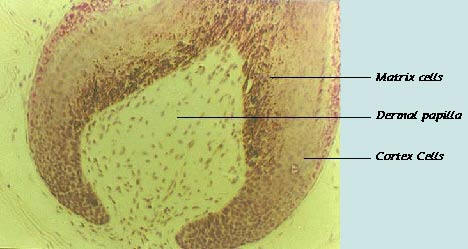
Dermal papilla - The dermal papilla directs and dictates the
embryonic
generation of a hair follicle and it also retains this instructive ability
throughout the life of the hair follicle. It consists of a highly active group
of cells shown to be capable of inducing follicle development from the
epidermis and production of hair fiber (Oliver 1966a, Oliver 1966b,
Oliver 1967).
The bigger the Dermal Papilla is, the more cells it has - the thicker the
hair fiber that the hair follicle produces.
Basement
Lamina - a thin layer of cells that separates the Dermal Papilla
form the hair sheath cells. Providing a barrier between the dermis and
epidermis.
Matrix
cells - Epidermal derived cells close to the Dermal Papilla. These
cells remain undifferentiated and focus on multiplying and proliferating
to produce more cells. Those cells made in the center of the hair follicle
are destined to become part of the hair fiber and are called Cortex
(cortical) cells.
Cortex
Cells - Cells that are made by the matrix cells, aimed to become a
part of the hair fiber.
As these cells
multiply, the constant stream of production pushes the cells
upward towards the skin surface. As they move up the hair follicle they
begin to differentiate into particular cell types. The cortex cells change
from a round into a flattened appearance. They are squeezed together into
layers (lamella). If the hair follicle contains melanocyte cells then
melanin pigment is incorporated into the cortex cells. These cortex cells
become keratinized and harden. As they do so it becomes impossible for
the cells to function properly and the cells die. The keratinized cells are
then pushed away from the hair bulb region and upwards as new cells
come in behind. The cortex cells are now part of the dead keratinized
fiber. Which is the hair that we see.
Capillaries
- small blood vessels that brings food and oxygen to the hair
follicles and takes away the CO2 and waste of the cells metabolic cycle.
When we give a
pulse of light, the light penetrates the epidermis and
absorbs in the cells.
The darker the
area the more energy it will absorb. As you can see in the
picture, the darkest area is the area of the matrix cells (the cells that
produce the hair fiber cells). That is because of the melanin, which is
inside these cells.
Another dark area is the Capillaries area - especially the capillaries that
have vain blood in them. Vain blood is darker because it has hemoglobin,
which is not attached to oxygen.
When we give a pulse of light many matrix cells are coagulated and stop
functioning.
Matrix cells that are not coagulated are triggered to activate their calcium
pumps. According to Friedmann (1993), a high intensity light pulse will
cause a vast calcium release from mitochondria into cytoplasm. The
hyperactivity of Ca+2 pumps exhausts the ATP (an energy molecule) pool
of the cell, thereby inhibiting cell metabolism resulting in the production
of less Cortex Cells and therefor - less hair or more feathery hair.
Another effect of the light pulse is the clotting of the vain blood in the
capillaries. The clotting disturbs the blood flow to the hair follicle
meaning less food and oxygen are delivered to the hair follicle, resulting
in a smaller ability to produce hair.
These three effects are the cause of the hair removal. The coagulation is
the main reason but the other two effects contribute to the hair loss as
well.
The Crystal 512 and Record 618delivers enough light energy, in the right wavelength, in
order to cause these effects but avoids from too much energy that might
influence other areas of the skin.